
LIVER CANCER & TUMOURS · SURGICAL ONCOLOGY
Liver cancer & tumours — expert surgery, explained honestly
A liver tumour diagnosis can feel overwhelming — but many liver tumours can be removed with intent to cure, and surgery in the right hands makes all the difference. Dr Ameet Kumar is an AIIMS-trained HPB & GI-oncology surgeon, FRCS (Glasgow), with 4,500+ surgeries across 25 years, who will tell you clearly where you stand.
M.Ch (GI Surgery) AIIMS New Delhi | FRCS (Glasgow) | FAIS | 4,500+ Surgeries
4,500+
Surgeries
25+
Years Experience
56
Publications
FRCS (Glasgow)
AIIMS-trained GI & HPB Surgeon
What liver cancer & tumours are
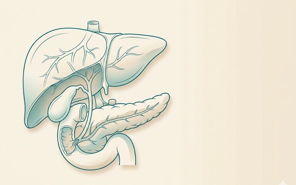
The liver is the body’s largest internal organ, involved in hundreds of metabolic functions. Tumours within it fall into two broad groups:
Primary liver cancer
This is cancer that starts in the liver itself. The most common type is hepatocellular carcinoma (HCC), which typically arises on a background of liver cirrhosis or chronic hepatitis B or C infection. HCC can grow slowly enough that, if caught on surveillance, surgery offers a genuine chance of cure. Less common primary tumours include cholangiocarcinoma (arising from bile ducts within the liver) and hepatic angiosarcoma.
Secondary liver tumours (metastases)
These are tumours that have spread to the liver from a cancer elsewhere in the body — most commonly colorectal (bowel) cancer. Colorectal liver metastases are, in the right patient, surgically curable. Other sources include breast, neuroendocrine tumours, and stomach cancer. The liver is a common site for spread, but “spread to the liver” does not automatically mean the situation is beyond surgery.
Benign liver tumours
Haemangiomas (benign blood vessel tangles) and focal nodular hyperplasia (FNH) are common incidental findings on imaging; most need only monitoring. Hepatic adenomas, particularly large ones, may warrant surgery due to a small risk of rupture or malignant change.
Symptoms & red flags
Primary liver cancer is often silent in its early stages, which is why regular surveillance in high-risk patients is so important. When symptoms do appear, they may include:
- Pain or fullness in the upper-right abdomen — a dull ache or sense of pressure under the right ribs
- Unexplained weight loss — significant and progressive
- Jaundice — yellowing of the skin or whites of the eyes
- Loss of appetite and persistent fatigue
- A palpable lump in the upper abdomen
- Worsening liver function in a patient already known to have cirrhosis or hepatitis
Patients with known cirrhosis or chronic hepatitis B or C should be on a formal 6-monthly ultrasound surveillance programme — HCC detected at this stage is often small and potentially curable.
Causes & risk factors
For primary liver cancer (HCC), the most significant risk factors are:
- Chronic hepatitis B or C infection — the commonest underlying cause worldwide
- Liver cirrhosis — from any cause, including alcohol, hepatitis, or non-alcoholic fatty liver disease (NAFLD)
- Non-alcoholic fatty liver disease (NAFLD/NASH) — increasingly common; can progress to cirrhosis even without heavy alcohol use
- Heavy, long-term alcohol use
- Aflatoxin exposure — a naturally occurring mould toxin found in some foodstuffs, particularly in tropical regions
- Family history — first-degree relatives with HCC or hereditary liver conditions
For secondary liver tumours (metastases), the risk factor is having an existing primary cancer elsewhere — most commonly colorectal (bowel) cancer, which frequently spreads to the liver via the portal circulation. Staging scans after a bowel cancer diagnosis routinely assess the liver for this reason.
How liver cancer is diagnosed
An accurate diagnosis requires careful imaging and, in most cases, specialist multi-disciplinary discussion before any treatment decision is made. The assessment typically includes:
- Ultrasound — usually the first-line test; identifies a lesion and prompts further imaging
- Triple-phase CT scan or MRI with contrast — the gold standard for characterising liver lesions; the enhancement pattern during different vascular phases is often diagnostic for HCC without the need for biopsy
- AFP (alpha-fetoprotein) — a blood marker that is elevated in many (though not all) HCC cases; useful alongside imaging
- Liver biopsy — not always required; used selectively when imaging is indeterminate or to confirm the primary site for metastatic disease
- Assessment of liver function — essential before any surgery; tests include Child-Pugh score, MELD score, and volumetric assessment of the future liver remnant (FLR)
- Multidisciplinary tumour board (MDT) — every case should be reviewed by a team including HPB surgeon, hepatologist, oncologist, and radiologist
Treatment options
The right treatment depends on the type of tumour, its size and location, the number of lesions, and the underlying health of the liver. Options include:
Liver resection (surgical removal)
Surgery remains the best curative treatment for both primary liver cancer and resectable colorectal liver metastases. The extent of resection is tailored to the tumour — a segmentectomy removes one segment of the liver, while a hemihepatectomy removes an entire lobe (left or right). The liver has a remarkable ability to regenerate, so large resections are possible provided adequate liver function is preserved.
Minimally invasive (laparoscopic) liver resection
Where the tumour location and patient fitness allow, keyhole (laparoscopic) liver resection offers the same oncological outcome with less blood loss, a shorter hospital stay, and faster recovery. Dr Kumar performs laparoscopic liver resections in suitable cases.
Ablation
For small tumours (typically under 3 cm), radiofrequency ablation (RFA) or microwave ablation can destroy the lesion using heat delivered via a needle under imaging guidance. Ablation is sometimes used alongside surgery or in patients who cannot undergo resection.
Combined approach for colorectal liver metastases
When bowel cancer has spread to the liver, treatment often involves chemotherapy combined with surgery. Some patients require downstaging — shrinking the tumour with chemotherapy first to make resection possible. The MDT reviews timing and sequencing of surgery on the bowel and the liver, which may be simultaneous or staged.
Liver transplant referral
In selected patients with HCC within strict criteria (the Milan criteria) on a background of cirrhosis, liver transplantation offers both a cure for the cancer and replacement of the diseased liver. Where appropriate, Dr Kumar will refer to a transplant centre and support the process.
How Dr Kumar treats liver cancer
Dr Ameet Kumar is a high-volume HPB surgeon trained at AIIMS New Delhi with an FRCS (Glasgow) — specialising in complex liver, pancreatic, and biliary surgery. His approach to liver cancer combines technical expertise with honest, patient-centred communication:
- Careful volumetric assessment — calculating the future liver remnant (FLR) before surgery to ensure sufficient liver remains post-resection
- Patient selection — matching the right operation to the right patient, considering both tumour biology and underlying liver health
- Keyhole liver resection where suitable — laparoscopic approach for appropriate tumour locations, reducing recovery time
- MDT-driven planning — every complex case reviewed with hepatology, oncology, and radiology before any decision
- Honest counselling — clear explanation of what surgery can and cannot achieve; no false promises, no alarm
He consults at Sakra World Hospital, Bengaluru, and sees patients from across India for second opinions, complex resections, and multidisciplinary planning.
da Vinci Robotic Surgery
Now also available with robotic assistance
Dr Kumar now also offers liver resection (hepatectomy) using the da Vinci robotic surgical system, for suitable tumour locations. Its 3D magnified vision and wristed instruments help with precise, controlled division of liver tissue, and the potential benefits include less blood loss, less pain and a shorter hospital stay. Open, laparoscopic and robotic approaches are all available — Dr Kumar will recommend whichever gives you the safest, most complete result for your case.
Recovery & outcomes after liver surgery
The liver has a unique capacity to regenerate — even after removing a significant portion, the remaining liver grows back to near its original volume over several weeks. This is what makes large liver resections safe in patients with a healthy liver. Key points about recovery:
- Hospital stay — typically 3–7 days for laparoscopic resection, 5–10 days for open surgery, depending on the extent of the resection
- Return to normal activity — most patients are back to light activities within 4–6 weeks
- Follow-up imaging — regular CT or MRI scans in the months and years after surgery to monitor for any recurrence
- Liver function monitoring — blood tests to track liver recovery in the early post-operative period
For patients with underlying cirrhosis, recovery requires closer monitoring as the remaining liver has less reserve. The pre-operative assessment specifically plans for this.
Frequently asked questions
Primary liver cancer starts in the liver itself — the most common type is hepatocellular carcinoma (HCC), which arises from liver cells, usually in a setting of cirrhosis or chronic hepatitis. Secondary liver cancer (metastases) has spread to the liver from a cancer that began elsewhere in the body, most commonly the bowel (colorectal cancer). The treatment approach differs for each, though surgery can be curative for both in selected patients.
Yes — many cases of liver cancer are surgically resectable, and surgery offers the best chance of a curative outcome. Resectability depends on the size, location, and number of tumours, the underlying health of the liver, and whether the disease has spread beyond the liver. Not every scan that looks complex is inoperable — a specialist assessment is essential before any conclusion is drawn.
In a patient with a healthy liver (no cirrhosis), up to 70–75% of the liver volume can be removed safely, because the remaining liver regenerates. In patients with underlying cirrhosis or hepatitis, less can be removed safely because the diseased liver has reduced regenerative capacity. A volumetric assessment (measuring the future liver remnant on CT) is performed before all major resections to ensure the operation is safe.
Yes — laparoscopic (keyhole) liver resection is feasible for many tumours, particularly those in the outer segments of the left lobe or anterolateral segments of the right lobe. Not all liver resections are suitable for a keyhole approach; the decision depends on tumour location, size, and proximity to major blood vessels and bile ducts. Where a laparoscopic approach is safe and appropriate, it offers less blood loss, shorter hospital stay, and faster return to activity.
Cirrhosis does not automatically rule out surgery, but it significantly changes the assessment. Liver function tests, the Child-Pugh score, and volumetric planning all form part of a careful workup. Patients with well-compensated cirrhosis (Child-Pugh A) and a small tumour in a favourable location may still be good surgical candidates. For those with more advanced cirrhosis and early HCC meeting strict criteria, liver transplantation — rather than resection — may be the preferred curative option. The MDT review is essential.
Yes — this is one of the most important messages in GI oncology. Colorectal liver metastases are not automatically incurable. In selected patients, surgical removal of both the bowel primary and the liver metastases — often combined with chemotherapy — gives a genuine prospect of long-term disease-free survival. Some patients require downstaging chemotherapy first to make the liver disease operable. The key is having the case reviewed by a specialist HPB surgeon experienced in colorectal liver metastasis surgery.
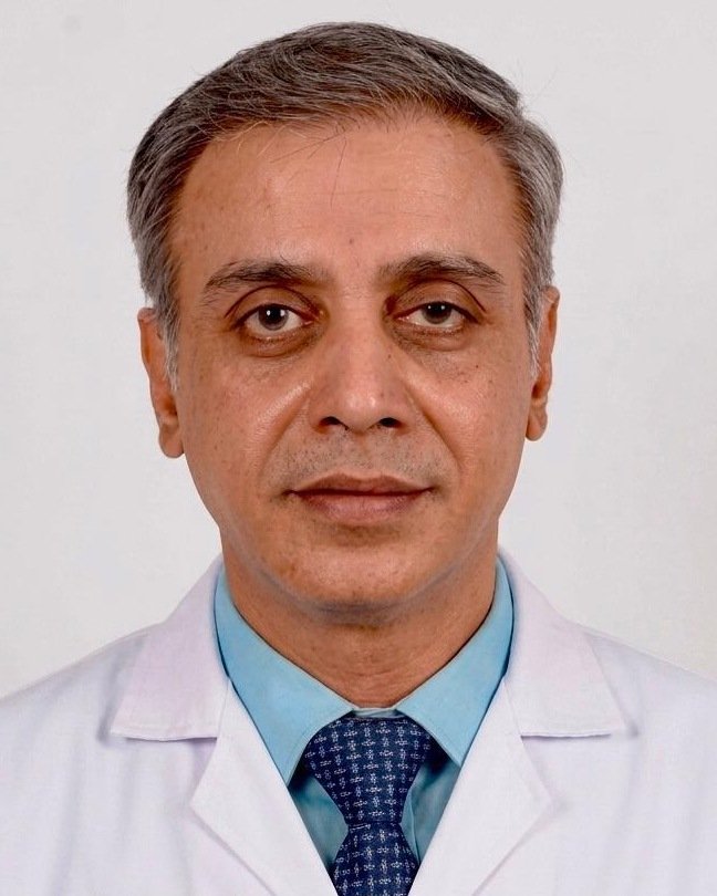
Written & medically reviewed by Dr Ameet Kumar, M.Ch (GI Surgery, AIIMS), FRCS (Glasgow), FAIS. Last updated: June 2026.
A liver tumour or liver cancer diagnosis? Let’s look at your scans together.
Whether you have a new diagnosis, a report that says “inoperable”, or imaging that needs expert interpretation — Dr Kumar will review your case and tell you honestly what surgery can offer. Early assessment makes all the difference. Book a consultation or message his team on WhatsApp.
Call 9606028116