
OESOPHAGEAL (FOOD-PIPE) CANCER · SURGICAL ONCOLOGY
Oesophageal (food-pipe) cancer — expert surgery, explained clearly
An oesophageal cancer diagnosis is frightening. When found in time, it can be treated with intent to cure. Dr Ameet Kumar is an AIIMS-trained GI-oncology surgeon, FRCS (Glasgow), with 4,500+ surgeries across 25 years — he will assess your case carefully and explain every option in plain language.
M.Ch (GI Surgery) AIIMS New Delhi | FRCS (Glasgow) | FAIS | 4,500+ Surgeries
4,500+
Surgeries
25+
Years Experience
56
Publications
FRCS (Glasgow)
AIIMS-trained GI & HPB Surgeon
What oesophageal (food-pipe) cancer is
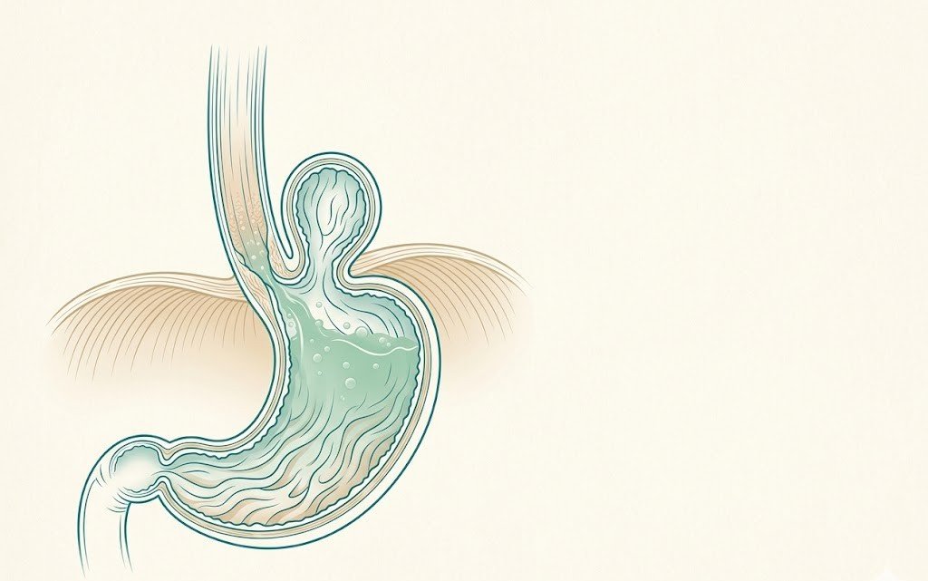
The oesophagus is the muscular tube — your food pipe — that carries food and liquid from the mouth to the stomach. It is about 25 cm long and runs through the chest. When cells lining the oesophagus grow abnormally and invade surrounding tissue, the result is oesophageal cancer.
There are two main types:
- Adenocarcinoma — the most common type in India today; arises from glandular cells typically in the lower oesophagus and gastro-oesophageal junction. Strongly linked to long-standing acid reflux (GERD) and Barrett’s oesophagus. Rates are rising globally.
- Squamous cell carcinoma — arises from the flat cells lining the upper and mid oesophagus; historically more common in South Asia; linked to smoking, alcohol and hot beverage consumption.
The oesophagus has no serosal (outer protective) layer, so tumours can spread relatively early to surrounding structures and lymph nodes. This makes careful staging and timely surgical assessment important. When the disease is caught while localised, curative surgery — an oesophagectomy — offers the best chance of long-term control.
Symptoms & red flags
Oesophageal cancer often has no symptoms in its earliest, most treatable stage. As the tumour grows it begins to narrow the food-pipe. The most important warning signs are:
- Progressive difficulty swallowing (dysphagia) — starting with solids (bread, meat) and advancing to soft food, then liquids. This is the cardinal symptom and demands prompt endoscopy.
- Food sticking or getting stuck — a sensation that food is lodging behind the breastbone or in the throat.
- Unintentional weight loss — significant, unexplained loss of weight over weeks to months.
- Persistent or worsening acid reflux / heartburn — especially when it is long-standing or has changed in character.
- Painful swallowing (odynophagia) — a burning or squeezing sensation when swallowing.
- Regurgitation of food — bringing up undigested food without nausea.
- Persistent hoarseness or cough — when a tumour affects nerves near the voice box.
If you have been diagnosed with Barrett’s oesophagus — a pre-cancerous change in the lower food-pipe lining caused by longstanding reflux — regular surveillance endoscopy is essential even if you feel well. Any new difficulty swallowing or change in reflux symptoms in someone with Barrett’s warrants an urgent review.
Causes & risk factors
Several factors are known to increase the risk of developing oesophageal cancer:
- Chronic acid reflux (GERD) and Barrett’s oesophagus — the leading risk factor for adenocarcinoma; longstanding, uncontrolled reflux damages the lower oesophageal lining, leading to Barrett’s, which can progress to cancer over years.
- Smoking and tobacco use — a major risk factor for both cancer types; doubles or triples risk.
- Heavy alcohol consumption — particularly for squamous cell carcinoma; the combination of smoking and alcohol multiplies risk significantly.
- Obesity — increases intra-abdominal pressure, worsens reflux, and is independently associated with adenocarcinoma of the lower oesophagus.
- Consumption of very hot beverages — repeatedly drinking scalding liquids (above ~65℃) is a recognised risk factor for squamous cell carcinoma.
- Diet low in fruits and vegetables — a diet poor in antioxidants and micronutrients is associated with higher risk.
- Achalasia — a motility disorder causing food to pool in the oesophagus; raises long-term cancer risk if untreated.
- Age and sex — more common after age 60 and significantly more common in men than women.
Having one or more risk factors does not mean you will develop cancer — but it does mean you should discuss surveillance and lifestyle modification with your doctor.
How oesophageal cancer is diagnosed
Diagnosis and staging follow a structured sequence. Every investigation has a purpose — together they build a picture of exactly where the tumour is, what type it is, and whether it has spread:
- Upper GI endoscopy (OGD scope) with biopsy — the first and essential investigation; the endoscopist visualises the tumour and takes small tissue samples to confirm the cancer type.
- Endoscopic ultrasound (EUS) — an endoscope with an ultrasound probe assesses how deeply the tumour has penetrated the oesophageal wall (T-staging) and samples nearby lymph nodes.
- CT scan of chest, abdomen and pelvis — assesses spread to lymph nodes and distant organs (liver, lungs, adrenals).
- PET-CT scan — identifies metabolically active cancer deposits that CT may miss; particularly important before planning surgery.
- Staging laparoscopy — for lower oesophageal and gastro-oesophageal junction tumours, a brief keyhole look inside the abdomen can detect small peritoneal deposits invisible on cross-sectional imaging.
- MDT / tumour board review — all findings are presented to a multidisciplinary team (gastroenterologist, oncologist, surgeon, radiologist, pathologist) to agree on the optimal treatment plan.
Accurate staging is not a delay — it is what makes the difference between the right treatment and the wrong one.
Treatment options
Treatment depends on the stage of the cancer, its location in the oesophagus, the type (adenocarcinoma or squamous cell), and the patient’s overall fitness. For localised disease, surgery remains central to curative treatment.
Surgery: oesophagectomy
An oesophagectomy removes the tumour-bearing segment of the oesophagus along with surrounding lymph nodes, and uses the stomach (or occasionally a segment of colon) to reconstruct the food passage. The main approaches are:
- Ivor Lewis oesophagectomy — an abdominal incision to mobilise the stomach, followed by a right thoracic (chest) incision to remove the tumour and join the stomach to the remaining oesophagus in the chest; used for mid and lower oesophageal tumours.
- McKeown (three-stage) oesophagectomy — adds a neck incision so the join (anastomosis) is made in the neck; used for upper oesophageal tumours.
- Minimally invasive oesophagectomy (MIO) — laparoscopic (keyhole) abdominal phase combined with thoracoscopic (keyhole chest) approach; when the tumour and patient fitness allow, this reduces the surgical trauma of the open technique, with shorter hospital stay and faster recovery.
Neoadjuvant chemotherapy or chemoradiotherapy before surgery
For many patients with localised but more advanced tumours, the MDT recommends chemotherapy — or combined chemotherapy and radiotherapy — given before surgery. This can shrink the tumour (downstage it), reduce microscopic spread, and improve the chances of a clear surgical margin. After completing treatment, re-staging scans determine whether surgery proceeds. This sequenced approach is now the standard of care for many oesophageal cancers.
Endoscopic resection for very early disease
When a cancer is detected at a very early stage — confined entirely to the innermost lining of the oesophagus — it may be removed endoscopically (through the endoscope, without open surgery) using techniques such as endoscopic mucosal resection (EMR) or endoscopic submucosal dissection (ESD). This is only appropriate for superficial, well-defined lesions, and the decision rests on careful EUS staging and MDT discussion.
Palliative treatments for advanced disease
When the disease is too advanced for curative surgery, the focus shifts to maintaining quality of life. Options include oesophageal stenting (a metal stent placed endoscopically to keep the food-pipe open), palliative chemotherapy, radiotherapy for symptom control, and nutritional support. Dr Kumar works closely with oncology and palliative care colleagues to ensure every patient receives coordinated, compassionate care regardless of stage.
How Dr Ameet Kumar approaches oesophageal cancer
Dr Ameet Kumar is a high-volume GI-oncology and HPB surgeon trained at AIIMS New Delhi — India’s premier surgical institution — and holds the FRCS qualification from the Royal College of Surgeons of Glasgow. With 4,500+ surgeries across 25 years, including oesophagectomies in complex patients, he brings both technical precision and honest, unhurried communication to every consultation.
His approach to oesophageal cancer:
- Thorough staging before any decision — reviewing all imaging, pathology, and EUS findings to establish exactly what surgery can achieve.
- MDT planning — working with oncologists and radiologists to sequence any neoadjuvant treatment correctly before operating.
- Minimally invasive oesophagectomy where appropriate — laparoscopic and thoracoscopic techniques when tumour location, staging, and patient fitness allow; smaller incisions, faster recovery, lower pulmonary complication rates.
- Ivor Lewis or McKeown oesophagectomy when open surgery is indicated — performed to oncological principles with careful lymph node clearance.
- Honest counselling — Dr Kumar will tell you clearly what the operation involves, what the realistic expectations for recovery are, and where surgery is not the right answer.
He is a Consultant at Sakra World Hospital, Bengaluru, working with a multidisciplinary GI oncology team to offer surgical care to the standard he developed over a 25-year career at tertiary military and teaching hospitals.
da Vinci Robotic Surgery
Now also available with robotic assistance
Dr Kumar now also offers oesophagectomy (food-pipe cancer surgery) using the da Vinci robotic surgical system, for selected patients. Its 3D magnified vision and wristed instruments allow precise dissection and lymph-node clearance in the tight spaces of the chest and abdomen, with the potential for less blood loss and a faster recovery. Open, laparoscopic and robotic approaches are all available — Dr Kumar will recommend whichever gives you the safest, most complete result for your case.
Recovery & what to expect after an oesophagectomy
An oesophagectomy is a major operation and recovery is gradual, but most patients are able to resume a good quality of life. Here is what the journey typically looks like:
- Hospital stay — typically 7–14 days, including time in a high-dependency unit for the first day or two. Most patients begin sips of water within 2–3 days of surgery.
- Eating again — food intake is reintroduced gradually: clear liquids, then soft foods, then a more normal diet over several weeks. The key principle is small, frequent meals — the reconstructed stomach is smaller and empties differently.
- Dietary adjustments — avoiding large meals, lying down immediately after eating, and very hot or very cold food in the first weeks; a dietitian helps plan appropriate nutrition. Some patients experience “dumping syndrome” (dizziness or diarrhoea after eating) which usually improves over months.
- Nutritional support — a jejunal feeding tube is often placed at the time of surgery to maintain nutrition during the early recovery phase; this is temporary.
- Return to activity — gentle walking from day one; most patients are back to light daily activities within 4–6 weeks. Heavier work takes longer.
- Follow-up — regular clinical review, CT scans, and endoscopy as per oncology protocols to monitor for recurrence and manage any long-term symptoms.
Recovery is a team effort. Dr Kumar and the multidisciplinary team will guide you through every step — from the operation through to nutritional rehab and long-term surveillance.
Frequently asked questions
No — difficulty swallowing (dysphagia) has many causes, most of which are benign. A narrowing or stricture from longstanding acid reflux, achalasia (a motility disorder), a pharyngeal pouch, or even anxiety can all cause food to feel like it is sticking. However, progressive dysphagia — starting with solids and worsening to liquids — combined with weight loss is the pattern most associated with a structural cause and warrants an urgent endoscopy to exclude cancer. Do not wait and see if the symptom is progressing.
Yes — when oesophageal cancer is detected while it is still confined to the oesophagus (and ideally while localised), surgical resection with curative intent is possible. Early-stage cancers detected during Barrett’s surveillance, or cancers that respond well to neoadjuvant chemotherapy before surgery, carry the best outcomes. The key is prompt referral, accurate staging, and treatment at a centre with experience in oesophageal surgery. Do not assume from a report or from reading online that an operation is not possible — a specialist review is essential.
An oesophagectomy removes the tumour-bearing part of the oesophagus and uses the stomach (brought up into the chest or neck) to reconstruct the food passage. It is a major operation — typically 4–6 hours under general anaesthesia — and is performed through a combination of abdominal and chest (or neck) incisions, or by keyhole (minimally invasive) technique when appropriate. The operation requires a surgeon experienced in oesophageal surgery and is undertaken in a hospital with high-dependency or intensive care facilities. Recovery in hospital is typically 7–14 days.
In suitable patients, yes. Minimally invasive oesophagectomy (MIO) uses laparoscopic (abdominal keyhole) and thoracoscopic (chest keyhole) techniques to perform the same operation through small incisions rather than large open cuts. This is associated with less post-operative pain, fewer pulmonary (chest) complications, a shorter hospital stay, and faster return to normal activity compared to open surgery — while achieving the same oncological (cancer clearance) result. Whether MIO is appropriate depends on the tumour’s position and stage, the patient’s general health, and the surgeon’s experience with the technique. Dr Kumar will advise clearly which approach is most suitable for your case.
Most patients can eat a wide variety of foods after recovery, but eating habits change. Because the stomach is reshaped to form the new food passage, it holds less than before. The key adjustment is eating small, frequent meals (5–6 times a day) rather than three large ones. Avoiding eating within 2–3 hours of lying down reduces reflux. Some patients experience “dumping syndrome” early on — a feeling of flushing, dizziness, or loose stools shortly after eating — which usually improves considerably over 6–12 months as the body adapts. A dietitian will guide you through the dietary changes from the first weeks after surgery onwards.
Barrett’s oesophagus is a pre-cancerous change in the lining of the lower oesophagus caused by longstanding acid reflux. Normal squamous cells are replaced by intestinal-type cells that carry a higher — though still relatively small — risk of progressing to adenocarcinoma over years. Most patients with Barrett’s do not develop cancer, but they do need regular surveillance endoscopy (typically every 2–3 years, more often if dysplasia is found) so that any progression is caught early. Surgery is not routinely needed for Barrett’s itself — it is managed with acid suppression medication and endoscopic surveillance. If high-grade dysplasia or early cancer is found, endoscopic ablation (such as radiofrequency ablation) or endoscopic resection may be offered before cancer develops. Dr Kumar will review your Barrett’s reports and advise the appropriate surveillance or intervention plan.
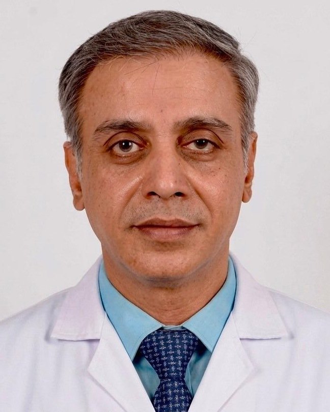
Written & medically reviewed by Dr Ameet Kumar, M.Ch (GI Surgery, AIIMS), FRCS (Glasgow), FAIS. Last updated: June 2026.
Trouble swallowing, or a new oesophageal diagnosis? Let’s review it together.
Whether you are newly diagnosed, waiting for a second opinion, or have imaging that needs expert surgical review — Dr Kumar will assess your case carefully and tell you honestly what surgery can offer. Early assessment matters. Book a consultation or message his team on WhatsApp.
Call 9606028116